Figure 4.
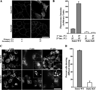
TGF-β receptor recycling is dependent on Dab2. (A) Direct recycling assay: Dab2 WT (top) and Dab2 KD16 cells stably expressing chimeric TGF-β receptors were incubated with mouse anti-GM-CSFR-β antibody for 1 h at 4 or 37°C (first incubation, left, top and bottom). Cells were subsequently washed and treated with acid to strip any surface, non-internalized antibody. Subsequently, cells were incubated with fluorescent Cy3-labeled anti-mouse secondary antibody for 1 h at 37°C (second incubation, right, top and bottom), washed, and acid treated again. This process allows only the receptors that have undergone 1.5 cycles of recycling to be fluorescently labeled (Mitchell et al., 2004). Cells were fixed, mounted, and quantified for cell associated fluorescence as described in Materials and Methods. (B) Data are represented as arbitrary units of fluorescence from 50 cells in each of three independent experiments from each clone. Images in A are from Dab2 KD16 and quantitation in B is an average of Dab2 KD16 and Dab2 KD18. (C) Transferrin recycling: Dab2 WT (top 3 panels) and Dab2 KD cells (bottom 3 panels) were incubated with Alexa Fluor 594 Tfn as described in Figure 2 for the indicated times. Cells were fixed and imaged as described in Figure 2. (D) Cells that sequestered Tfn internally in the perinuclear compartment and cells that showed diffused staining (indicative of Tfn recycling) at 15 min were counted manually. Data presented are expressed as percentage of diffused staining at 15 min seen in the WT cells and indicate the mean ± SD of 50 cells in each of three independent experiments. Images in C are from Dab2 KD16 and quantitation in D is an average of Dab2 KD16 and Dab2 KD18. Bar, 10 μm.

