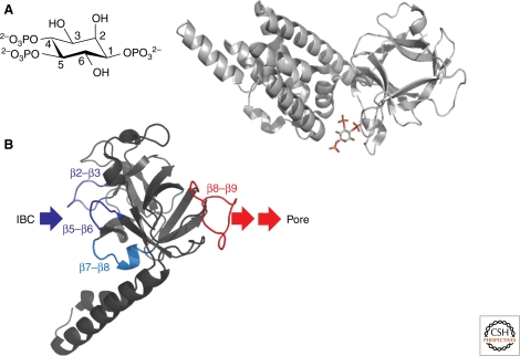
Figure 3.
Initiation of IP3R activation by IP3. (A) The structure of IP3, with its critical vicinal 4,5-bisphosphate and 6-hydroxyl groups, is shown alongside the structure of the IBC with IP3 bound. The latter shows the 4- and 5-phosphates contacting the β- and α-domains, respectively (Bosanac et al. 2002), and thereby pulling the clam into a more closed state. (B) Structure of the SD (Bosanac et al. 2005) showing possible sites of interaction with the IBC and downstream domains through which it signals to the pore. See text for further details.

