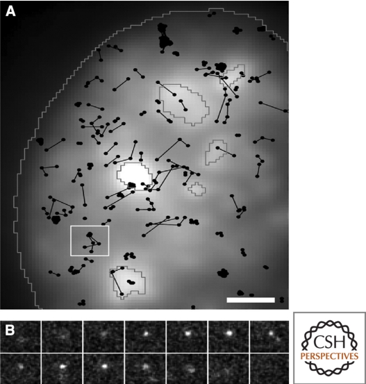
Figure 5.
In vivo trajectories of single U1 snRNPs within the nucleus of HeLa cells. Fluorescently labeled native U1 snRNPs were microinjected to visualize and track single molecules, recorded at 200 Hz. SF2/ASF-GFP was transiently expressed to distinguish mobile and transiently immobilized U1 snRNP particles within the nucleoplasm and speckles, outlined in gray (A). A 8 µm2 area from A is broken down into a short image sequence displaying a single trajectory over time (B). Grunwald et al. 2006, © 2006 by The American Society for Cell Biology.

