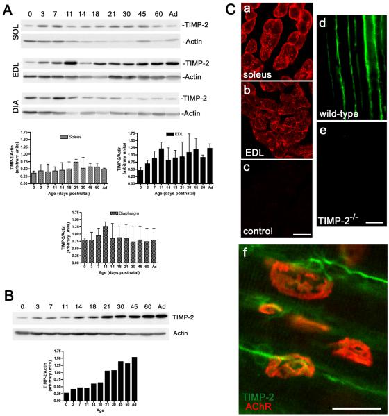
Figure 1. TIMP-2 is expressed at the neuromuscular junction.
A) Western blot analysis of soleus (SOL), extensor digitorum longus (EDL), and diaphragm (DIA) (25 μg crude homogenate) at the indicated postnatal ages (Ad-adult). TIMP-2 expression was normalized to actin as a loading control. TIMP-2 expression is greatest in EDL and is increased during development. B) Western blot analysis of spinal cord lumbar enlargement (20 μg crude homogenate). TIMP-2 is more abundantly expressed in spinal cord than muscle. Like the EDL, TIMP-2 expression in spinal cord is developmentally up-regulated. C) Confocal photomicrographs of TIMP-2 expression in transverse sections of P21 soleus (a, c), EDL (b), and longitudinal sections of P14 sartorius (d, e). Antibody specificity is demonstrated by the lack of immunolabeling with secondary antibody alone (c) and in TIMP-2−/− muscle (e). In addition to labeling muscle basal lamina, TIMP-2 (green) labels motor neurons at the NMJ endplate (red, rhodamine-conjugated α- BTX) (f). Scale bars = 25 μm.

