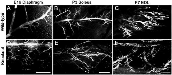
Figure 2. Intramuscular nerve branching is increased in TIMP-2−/− mice.
Confocal micrographs show neurofilament-145 immunolabeling in TIMP-2−/− mice and wild-type littermate controls. A well-defined central nerve trunk with branches emanating from it is present in wild-type E16 diaphragm (A) and P3 soleus (B), while the P7 EDL shows a discretely organized endplate band (C). This is in stark contrast to the nerve branching in TIMP-2−/− mice. The altered TIMP-2−/− nerve morphology may be due to increased nerve branching (D) and/or axon defasciculation (E); thereby, resulting in a disorganized endplate region (F). Altered nerve branching is also present in the diaphragm at P3, but not P7, and in the P3 EDL and P7 soleus (data not shown). Data are representative of 3 embryos and 6 neonatal mice. Scale bar = 100 μm.

