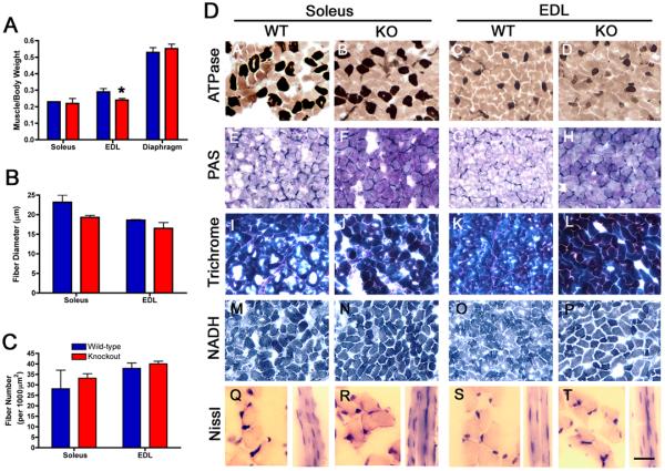
Figure 4. Muscle cytoarchitecture is normal in P21 TIMP-2−/− muscle.
A) Muscle weight, normalized to body weight, is reduced in TIMP-2−/− EDL (* p = 0.02), but not soleus (p = 0.5) or diaphragm (p = 0.5, n = 5). B) Fiber diameter is not reduced in soleus (wt: 21.4 ± 1.8, ko: 19.2 ± 0.5, p = 0.2) or EDL (wt: 18.4 ± 0.3, ko: 17.4 ± 1.2, p = 0.5, n = 3). C) The number of muscle fibers (per 10,000 μm2) is also not different in the soleus (wt: 28.2 ± 8.9, ko: 33.2 ± 2.2; p = 0.6) or EDL (wt: 37.9 ± 2.5, ko: 40 ± 1.3; p = 0.5). D) TIMP-2−/− muscles at P21 appear histologically normal; with the exception of increased basement membrane glycoproteins and collagen detected with Periodic Acid Schiff (PAS, E-H) and Masson Trichrome stain (I-L), respectively. ATPase at pH 4.3 shows no changes in slow-twitch muscle fiber number or distribution (A-D). In addition, no difference in NADH diaphorase is present (M-P). Examination of muscle cross sections reveals that TIMP-2−/− muscle lack central nuclei, but longitudinal sections appear to possess more nuclei per muscle fiber (Q-T). Scale bar = 50 μm (A-P), 25 μm (Q-T longitudinal sections), and 12.5 μm (Q-T cross sections).

