
Figure 6. Nudt16 is Involved in the Decay of a Subset of mRNAs.
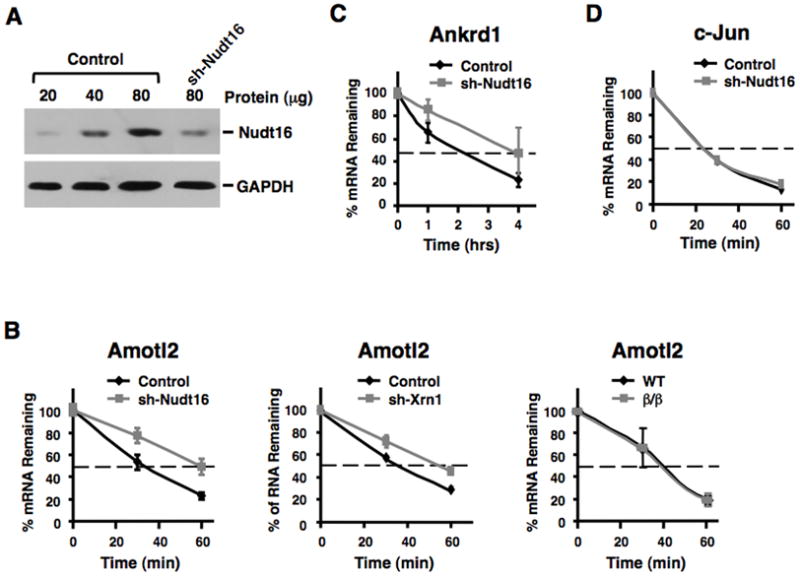
Amotl2 and Ankrd1 mRNAs are stabilized following a reduction in Nudt16 protein levels. (A) Nudt16 protein levels were detected following lentiviral expressed mNudt16-directed shRNA in MEF cells as determined by Western Blot analysis. (B) Stability of Amotl2 mRNA in control and Nudt16 knock down MEF cells are shown in the left panel and the fate of the same mRNA in wild type and Dcp2β/β MEF cells is shown on the right. The middle panel shows the decay of Amotl2 mRNA in cells with a lentiviral shRNA directed 65% reduction in Xrn1 protein. Transcription was arrested by the addition of Actinomycin D 48 hours post-lentiviral infection and mRNA levels tested at the times indicated. Quantitation of three independent experiments are shown with +/- standard deviation denoted by error bars. mRNA levels of Ankrd1 (C) and c-Jun (D) following transcriptional arrest are shown as described in B above.