
FIGURE 1. HE and FISH images of case 13.
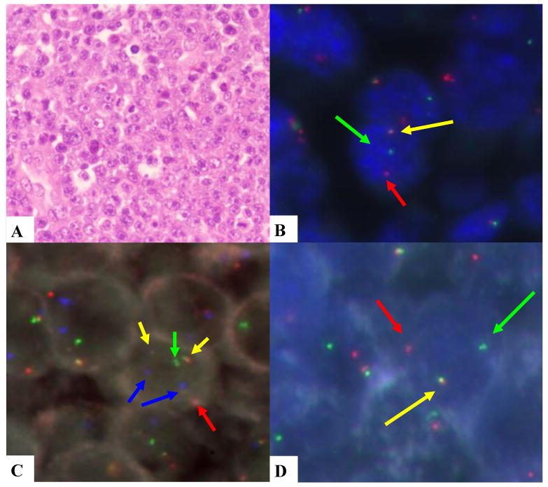
A. monomorphic PBL. B. MYC rearranged (break apart probe). C. IGH/MYC dual fusion probe; yellow signals indicate the IGH/MYC translocation, the green signal is the normal IGH locus, the red signal is the normal MYC locus and the blue signals are the CEP8. D. IGH rearranged (break apart probe).