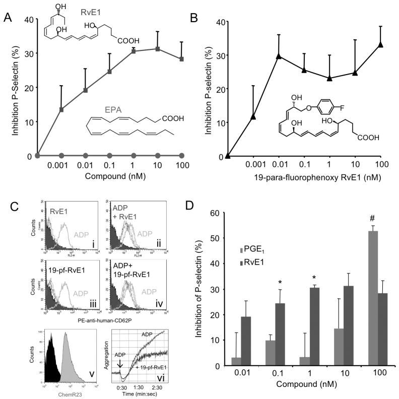
Figure 1. RvE1 reduces ADP-stimulated P-selectin surface mobilization.
PRP was incubated with either vehicle, (A) RvE1 (0.01–100 nM, blue), EPA (0.01–100 nM, red) (B) 19-para-flurorophenoxy-RvE1 (0.01–100 nM) was incubated for 15 minutes, 37°C, and ADP (10 μM) was added for 3 minutes, 37°C. Platelets were stained with PE-anti-human CD62P (P-selectin) and subjected to flow cytometry. (C) Representative histograms of PE anti-human CD62P surface expression. Vehicle (shaded), ADP (10μM) alone, solid line, RvE1 or 19-para-fluorophenoxy-RvE1 (dashed lines). (Ci) and (Ciii) indicate SPM alone while (Cii) and (Civ) display SPM + ADP. Histograms are representative of n=5–6 separate donors. (Cv) Representative histogram of ChemR23+ platelets. (Cvi) 19-para-fluourophenoxy-RvE1 (10 nM) reduction of ADP-stimulated platelet aggregation, representative of n=3. (D) Direct Comparison between PGE1 (0.01 nM-100 nM) and RvE1 at equi-molar concentrations. Results are mean ± SEM n=3, separate donors, *p<0.05, #p<0.03.

