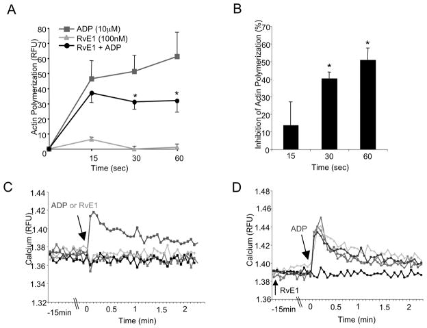
Figure 2. RvE1 reduces ADP-stimulated actin polymerization: Ca2+ independent.
(A,B) PRP was incubated with either vehicle or RvE1 (100 nM) for 15 minutes, 37°C with gentle mixing then stimulated with ADP (10 μM) for 0, 15, 30, or 60 seconds. Incubation was stopped by the addition of ice-cold formalin (6%) and platelets were permeabilized and stained with FITC-phalloidin (1:100) for 1 hour, 4°C. Changes in platelet morphology were quantified via flow cytometry and Cell Quest software. (B) RvE1 (100nM) percent inhibition of ADP-stimulated actin polymerization. Results are mean±SEM n=3, *p≤ 0.03. (C,D) Fluorescent intracellular calcium indicator, Fura-2. (C) RvE1 (1–100nM) as compared to vehicle and ADP. (D) RvE1 (1–100nM) plus ADP as compared to vehicle. Fluorescence was monitored by an EnVision plate reader and accompanying software. Results are representative of n=3.

