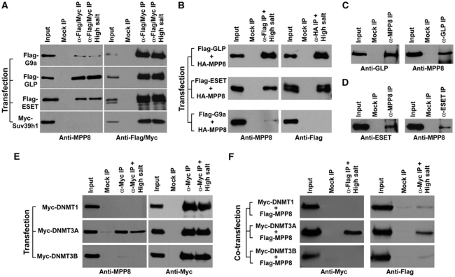
Figure 6.
MPP8 interacts with HMTase GLP, ESET and DNMT3A. (A) 293T cells were transfected with each of H3K9 HMTase expression vectors. Antibodies used for IP and following western blot are indicated. High salt represents washing buffer containing 600 mM KCl compared with normal (300 mM KCl). (B) Similar IP-western analysis after co-transfecting with expression vectors for different H3K9 HMTases together with HA-MPP8. Antibodies used for IPs and western blot are indicated. (C, D) MPP8 interacts with GLP and ESET endogenously. Cell extracts derived from 293T cells were incubated with anti-MPP8 and anti-GLP antibodies (C) or anti-MPP8 and anti-ESET antibodies (D) for IP. Endogenous protein complexes were analysed by western blot with antibodies indicated below each panel. (E) 293T cells were transfected with each of Myc-DNMT1, DNMT3A and DNMT3B expression vector and the similar IP-western analysis were carried out using indicated antibodies. High salt represents washing buffer containing 300 mM KCl compared with normal (150 mM KCl). (F) Co-transfections were performed with each of Myc-tagged DNMT vectors and Flag-MPP8 vector. IPs were carried out with under high salt condition and analysed by western blot using indicated antibodies. In all IP-western experiments, normal mouse or rabbit IgG was used for mock IPs and ‘Input' represents 5% of total cell extract.

