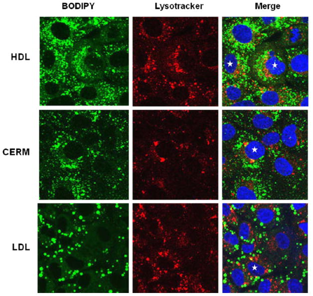
Figure 6.

Confocal fluorescence microscopy of the accumulation of Bodipy CE in Huh7 cells incubated with Bodipy-CE labeled HDL, CERM and LDL (Left panels, green). Cells were also stained with LysoTracker Red to label lysosomes (Middle panels, red) and with Hoeschst (blue) to label nuclei. The merged images (Right panels) show distinct green labeling of intracellular vesicles, as well as some co-localization with lysomes (yellow) as seen especially in the cells noted with the stars.
