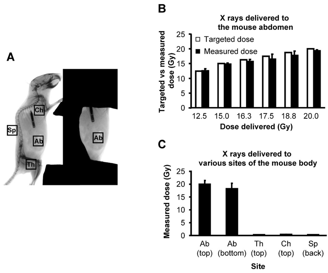
FIG. 2.
Verification of absorbed doses at the abdomen and validation of shielding efficacy. Ten-week-old male C57BL/6 mice under anesthesia were positioned for irradiation. After dosimetry performed using a PTW Farmer Chamber connected to a CNMC Electrometer that was positioned at the approximated mid-abdomen position, absorbed doses of X rays of various targeted doses at multiple body parts of a mouse were measured using the NanoDot dosimeter chips. Panel A: Positioning of the NanoDot dosimeter chips in a mouse exposed to radiation. NanoDot dosimeter chips were placed on the center of the top and bottom surface of the exposed abdomen (Ab) as well as on the top of the shielded left thigh (Th), chest wall (Ch) and spine (Sp) of anesthetized mice. The mouse was then exposed to 12.5–20.0 Gy X rays at a dose rate of 1.079 Gy/min using a Faxitron X-ray Generating System (CP-160, Faxitron X-Ray Corp., Wheeling, IL). Panel B: Comparison of targeted doses and absorbed doses measured on the top surface of the exposed mouse abdomen positioned for irradiation. Bars are the means and standard deviations calculated from readings on five mice, one reading per dose per mouse. Panel C: Absorbed doses measured at various sites of the mouse body positioned for irradiation. Data are the means and standard deviations calculated from readings on two mice with three readings per site per mouse.

