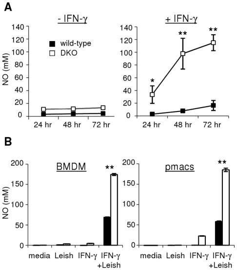
Figure 3. LXR-deficient macrophages produce increased levels of NO following exposure to Leishmania and IFN-γ in vitro.
A. BMDM from individual wild-type and DKO (Sv129xC57Bl/6) mice were stimulated in vitro with stationary-phase L. chagasi/infantum promastigotes for the times indicated, in the absence (left) or presence (right) of IFN-γ pre-treament. B. BMDM (left) or peritoneal macrophages (right) from wild-type (solid bars) or LXR-DKO (empty bars) mice (C57Bl/6 background) were infected with stationary-phase L. chagasi/infantum (with or without IFN-γ pre-treatment) for 24 hours in vitro. Supernatants were removed and levels of NO were quantitated by the Griess method. Error bars represent standard deviations of triplicate NO measurements. Results are representative of at least 3 independent experiments; * denotes p<0.05, ** p<0.005.

