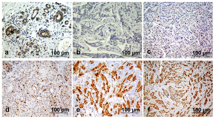
Figure 1.
Immunohistochemistry (IHC) analysis of ANCCA expression in normal or cancerous human breast tissues. Representative images are shown for IHC score 0 with less than 1% of nuclei stained positive, in a histologically normal breast tissue (a) and in a tumor (b); for score 1 with < 25% of positively stained nuclei in a tumor (c); for score 2 with 25–50% of positively stained nuclei a tumor (d); and for score 3 with >50% of positively stained nuclei in an ER-positive tumor (e) and an ER-negative tumor (f).

