
Figure 3.
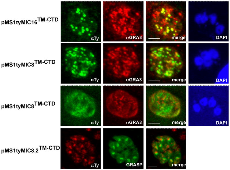
Localization of several stably transfected chimeras in the parasite using double immunofluorescence analysis and confocal microscopy. Anti-GRA3 or GRASP-YFP were used as dense granules and Golgi markers, respectively. Scale bars indicate 5μm. pMS1tyMIC16TM-CTD and pMS1tyMIC8TM-CTD expressing parasites were stained with anti-Ty-1 (in green) and anti-GRA3 (in red). pMS1tyMIC8.2TM-CTD was stained with anti-Ty-1 (in red) and co-localized with expression of GRASP-YFP (in green). The nucleus was stained with DAPI (in blue).