
Figure 5.
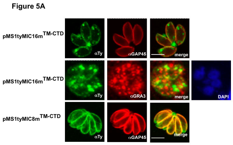
A Subcellular distribution of the SAG1-TM-CTD mutant chimeras by IFA and documented by confocal microscopy. Anti-GAP45 antibodies (in red), Anti-GRA3 (in red) and GRASP-YFP (in green) were used as IMC, DGs and Golgi markers, respectively. The nucleus was stained with DAPI (blue). pMS1tyMIC16mTM-CTD, pMS1tyMIC8mTM-CTD (in green) andpAS1tyAMA1mTM-CTD (in red) expressing parasites were stained with anti-Ty-1. Scale bars indicate 5μm.
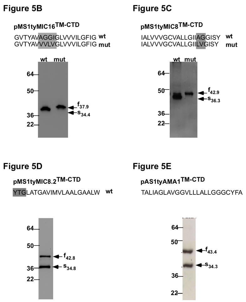
B–E Analysis of the cleavage events in the different chimeras. On the top, is shown the TMD of each TM-MIC in the original (wt) and mutated chimera (mut) and the residues mutated in the rhomboid cleavage motif are boxed. Below are western-blot analysis of lysates from parasites stably expressing the different pMS1tyMICTM-CTD fusion constructs. Indicated by arrows are the migration of the full (f) and shed (s) forms with indication of the molecular weight. B Migration of pMS1tyMIC16TM-CTD (wt) and pMS1tyMIC16mTM-CTD (mut). C Migration of pMS1tyMIC8TM-CTD (wt) and pMS1tyMIC8mTM-CTD (mut). D Migration of pMS1tyMIC8.2TM-CTD. E Migration of pAS1tyAMA1TM-CTD. The proteins were detected with anti-Ty-1. Molecular weight markers are indicated in kDa.