
Fig. 3.
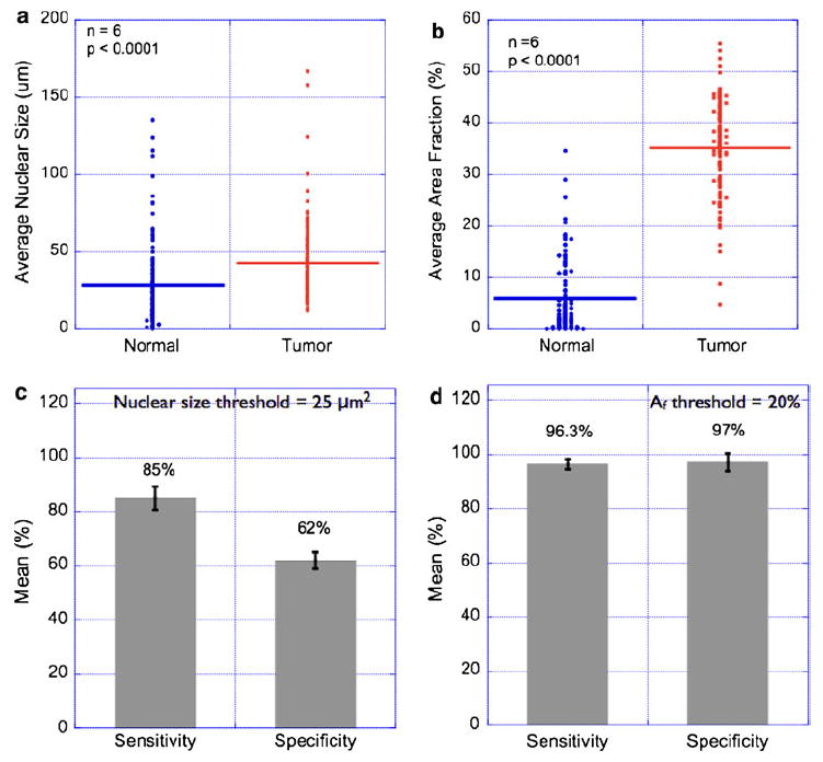
Statistical analysis of nuclear morphometry parameters in breast tissues. Nuclear morphometry parameters were calculated in multiple images of normal and breast-tumor specimens as described in the “Material and methods” section. Each image (462 μm × 346 μm size) was divided into sub-images of size (50 μm × 50 μm), and the mean nuclear size was computed. This step ensured that the entire image was sampled with uniform sampling interval. Thus, every data point in the Fig. 3a, b represents mean nuclear size in the predefined sub-image regions. Statistical data from six representative pairs of normal and tumor regions are presented in (a). As can be seen, the observed difference in mean nuclear size in the normal and tumor regions was found to be statistically significant. However, the estimated sensitivity and specificity values from these data were only 85 and 62%, respectively (c). In order to remedy this problem, we measured the nuclear area fraction (Af) which parameterizes a combination of nuclear size and number in a given region-of-interest. Statistical comparison of measured area fractions is shown in (b), and the corresponding sensitivity/specificity comparison is shown in (d). These results suggest that it is possible to achieve high sensitivity and high specificity in tumor diagnosis based on nuclear area fractions. It is worth mentioning that the nuclear size criterion can be made highly specific at the cost of decreasing sensitivity