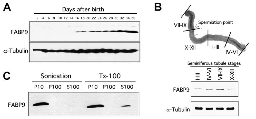
Fig. 2. Characterization of FABP9.
(A) FABP9 protein expression during post-natal testicular development. Immunoblot of testicular proteins collected every 2 days from mice aged day-2 to day-36 shows FABP9 protein expression starting at day-16 consistent with the appearance of pachytene spermatocytes. (B) Expression of FABP9 in different stages of germ cells in the seminiferous tubules. Highest expression was seen in tubular stages IV–VI and VII–IX which both contain germ cells in advanced stages of spermiogenesis. (C) FABP9 association with different sperm biochemical fractions separated by either physical shear (sonication) or detergent (TX-100). With sonication, FABP9 was found only in the P10 fraction that represents insoluble cytoskeletal structures and organelles. With TX-100, FABP9 was also seen predominantly in the P10 fraction although a small percentage solubilized and was found in the S100 fraction. FABP9 did not associate with the P100/membrane fraction.

