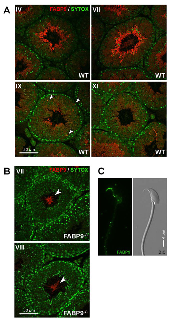
Fig. 5. Localization of FABP9 in developing male germ cells and sperm.
(A) FABP9 localization in testis sections. Seminiferous tubule cross-sections show FABP9 protein expression was abundant in late elongating spermatids. Earlier in male germ cell development, FABP9 localization was cytoplasmic and diffuse in spermatids. Labeling seen in the principal piece at advanced stages of spermiogenesis was deduced to be non-specific (based upon labeling observed in FABP9−/− testes). Arrowheads denote labeling of isolated, rare spermatocytes. (B) Specific labeling for FABP9 was absent in testis sections from FABP9−/− mice. Non-specific labeling was observed in the principal piece of testicular sperm (arrowheads). (C) FABP9 localization in mature sperm. In the permeabilized sperm head, FABP9 labeled the perforatorium intensely and the post-acrosomal peri-nuclear theca weakly. A corresponding DIC image is also shown.

