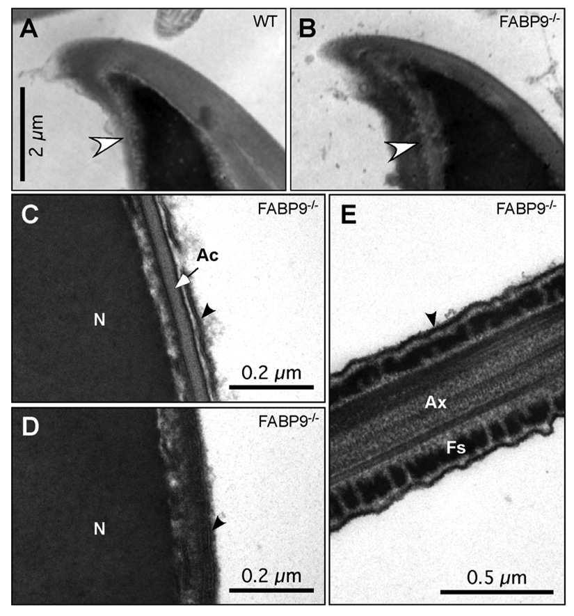
Fig. 8. Membrane tethering, and perforatorium and perinuclear theca morphology in FABP9−/− mice.
Ultrastructural examination of membrane attachment to the underlying structures revealed no abnormalities in FABP9−/− sperm (panels B–E). (A) The apical hook region of a WT sperm head. (B) The same region of a FABP9−/− sperm head is shown. Note that the two views are slightly oblique to one another, but both show the apical acrosome extending up the convex curve, both show the extension of the nucleus into the hook, and both show normal tethering of the acrosome to the underlying structure and normal tethering of the plasma membrane overlying the acrosome. Sperm ultrastructure and membrane tethering were also normal in null sperm in the equatorial region (C), the post-acrosomal region (D), as well as the principal piece (E). Black arrows point to the bilayer structure of the plasma membrane. [N: nucleus; Ac: acrosome; Ax: axoneme; Fs: fibrous sheath]

