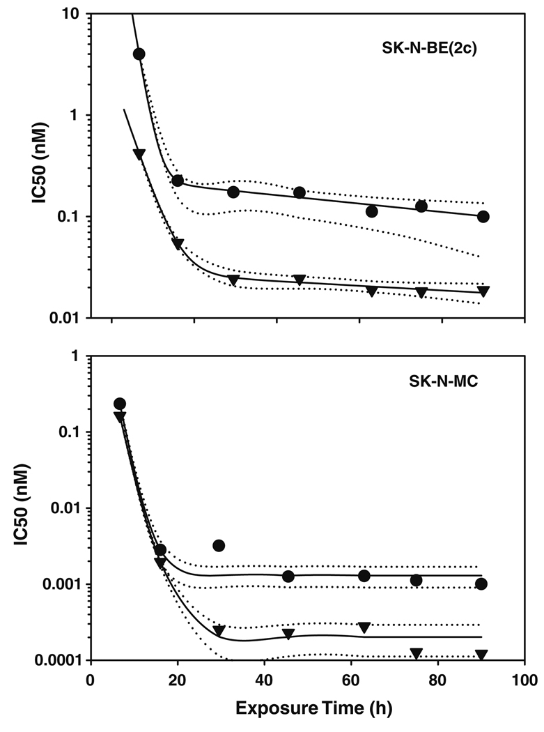
Fig. 1.
Activity of BACPT in neuroblastoma cells as a function of exposure time and extracellular pH. Neuroblastoma cell lines that do [SK-N-BE(2c)] or do not (SK-N-MC) over-express NET or MYCN were exposed to varying concentrations of BACPT for various times before being placed in drug-free medium prior to analysis of surviving cell number by ATP assay. The concentration that inhibited growth by 50% versus untreated controls (IC50) was determined as described in “Methods”. Cells were cultured at pH 7.4 (circles) or pH 6.8 (inverse triangles). Solid lines represent a curve fit of the data with dotted lines the 95% conidence intervals

