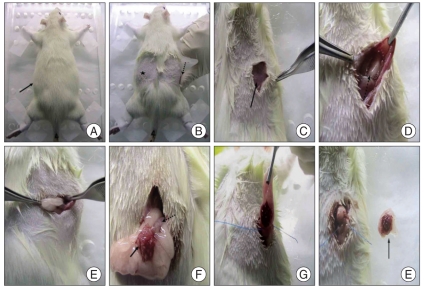
Fig. 2.
Procedures of ovariectomy in rat. Anesthetized rat is laid prone on operating table. Thick black arrow : shaving site (A). Skin incision point is located just medial portion of the most bulged area (★) or a thumb used to find the incision point (dotted arrow) (B). External oblique muscle is exposed after skin incision. Thick black arrow : External oblique muscle (C). After the muscle dissection, peritoneal space and adipose tissue surrounding ovary are exposed. Dotted arrow : adipose tissue surrounding ovary (D). The surrounding fat must be gently pulled to avoid detachment of small pieces of ovary (E). This shows ovary (thick black arrow) and uterine horn (dotted arrow) surrounded by fat (F). Ligation must undergo at distal uterine horn in order to get rid of total ovary at a time (G). Ovary surrounded by fat is removed totally (thick black arrow) (H).

