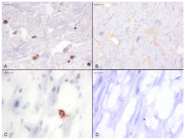
Fig. 2. Anti-Ucn 1 immunohistochemistry of canine ventricular myocardium.
Discreet foci of Ucn 1 positive staining are seen in ventricular (A) and atrial (C) tissue. No staining occurred in ventricular (B) and atrial (D) tissue incubated with control non-immune rabbit serum. Representative image, size bars are 10 μm, light haematoxylin counterstain.

