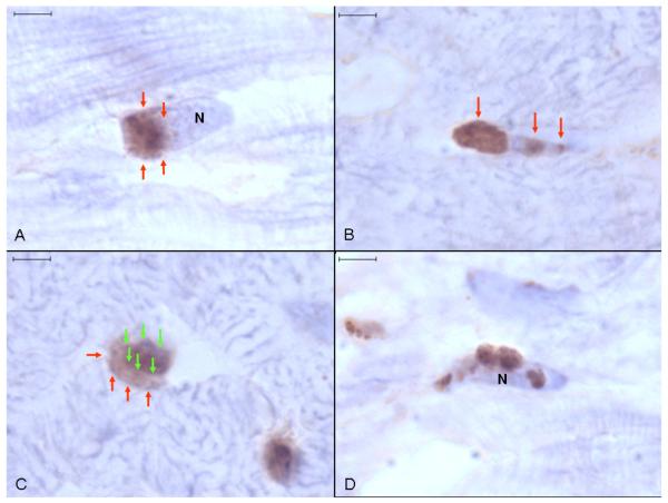
Fig. 3. Anti-Ucn 1 immunohistochemistry of canine ventricular myocardium.
(A) A round focus of Ucn 1-positive staining (arrows) adjacent to one pole of a haematoxylin stained cardiomyocyte nucleus. (B) Associated with a cardiomyocyte nucleus is a prominent focus of peripolar staining (left arrow) and several small bands of Ucn 1-positive staining perpendicular to the long axis of the nucleus (other arrows). (C) Ucn 1-positive staining (red arrows) extends beyond the boundaries of the cardiomyocyte nucleus (green arrows). (D) Irregular, random granular Ucn 1-positive staining appears to be adherent to a cardiomyocyte nucleus (N). Representative image, size bars are 4 μm, light haematoxylin counterstain.

