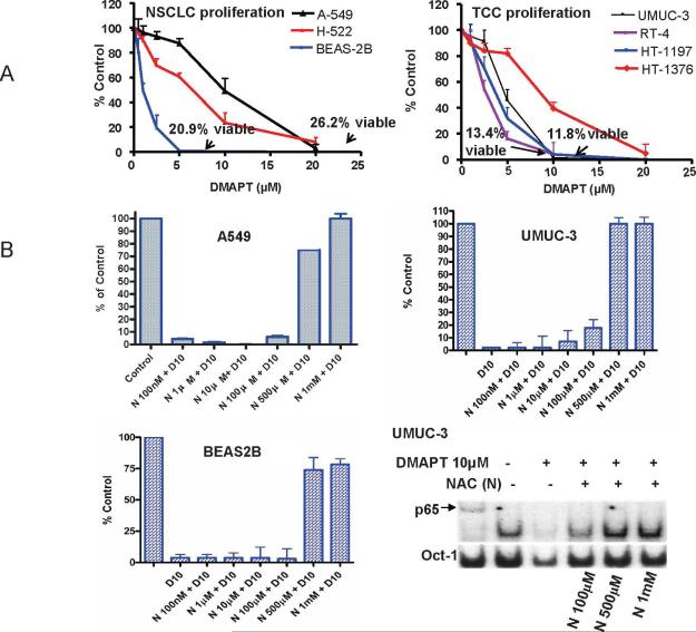
Figure 2. DMAPT inhibits cellular proliferation of early and late neoplastic lung and urothelial cell lines.
(A) BrdU incorporation assay demonstrated DMAPT's ability to inhibit the in vitro proliferation of NSCLC cell lines – A549, H-222 and BEAS (left panel) and TCC cell lines - UMUC, HT1197, HT1376 and RT-4 (right panel). For each cell line 2,000 cells were placed in 100μL, drug added at concentrations of 1, 2, 5, 10 and 20 μM after 24 hours of cell growth and the percentage of cells relative to untreated control was determined after 48 hours of drug exposure. The trypan blue assay showed that after one DMAPT dose the viability for each cell line at 48 hours was 26.2% for A549 with 20 M, 20.9% for BEAS2B with 5 M; 11.8% for UMUC-3 with 10μM and 13.4% for RT4 with 10uM. (B) Using a BrdU incorporation assay NAC was observed to decrease DMAPT's in vitro anti-proliferative activity in UMUC-3, A549 and BEAS2B in a dose dependent manner. In all cell lines, 10 M DMAPT suppressed proliferation by greater than 90% and this was almost completely abrogated by 1mM NAC in UMUC-3 and A549 and DMAPT's anti-proliferative efficacy was limited to only 25% in BEAS2B cell line. (B, bottom right panel). Electrophoretic mobility gel shift assay demonstrating NAC abrogating DMAPT's ability to inhibit NFκB DNA binding in a dose dependent manner in UMUC-3.

