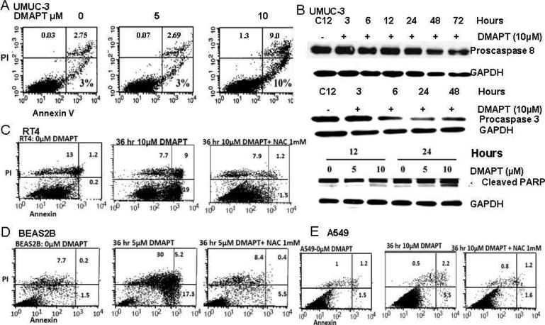
Figure 4. DMAPT induces cell death in a cell dependent manner.
(A) Flow cytometry with propidium iodine and Annexin V staining showed the ability of DMAPT to induce early and late apoptosis in UMUC-3 with 5 and 10 M. In this assay, the upper left quadrant with predominant PI staining represents necrosis, the upper right, combined PI and annexin staining represents late/atypical apoptosis and bottom right with predominant annexin staining represents early/typical apoptosis. (B) Western blotting of proteins in UMUC-3 detailed DMAPT induced cleavage of procaspase 8, procaspase 3, and PARP. Flow cytometry demonstrated DMAPT induced early and late apoptosis in RT4 (C), BEAS-2B (D) and A549 (E). In BEAS-2B, DMAPT also induced substantial necrosis. All of these types of cell death were blocked by NAC in RT4, BEAS2B and A549.

