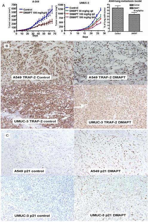
Figure 6. DMAPT has in vivo anti-cancer activity in both transitional cell and non-small lung cancers.
(A) Established xenograft experiments with A549 (left panel) and UMUC-3 (middle panel) cells were conducted with the cell lines injected into the subcutaneous tissue of the flanks of athymic male nude mice. Treatment was commenced when tumors were palpable at day 7. DMAPT was given by daily oral gavage and was able to suppress tumor growth relative to untreated control in A549. In UMUC-3, DMAPT's ability to suppress tumor growth was observed to be dose dependent with 100 mg/kg twice per day (bid) being more effective than 50 mg/kg or 100 mg/kg daily (qd). The ability of orally administered DMAPT to inhibit the in vivo metastatic process (implantation of circulating cells) of A549 cell lines (right panel) was demonstrated by measuring the relative volume of lung involved with cancer 60 days after cells were injected by tail vein. Treatment was commenced on day 1 with daily oral DMAPT given by oral gavage for 48 days. (B) TRAF-2 immunohistochemical staining of tumors is depicted by the brown staining of cells. The tumors were extracted from the control and DMAPT single agent arms at the time of sacrifice of both A549 and UMUC-3 experiments. The photographs of the microscopic images demonstrate that compared with control, DMAPT decreased the expression of TRAF-2 in both A549 (top panel) and UMUC-3 (bottom panel) as evidenced by decreased intensity of brown staining of cytoplasm in both cell lines as well as in the nuclei of UMUC-3 cells. (C) p21 immunohistochemical staining of tumors is depicted by brown staining of cells. The tumors were extracted from the control and DMAPT single agent arms at the time of sacrifice of both A549 and UMUC-3 experiments. The photographs of the microscopic images demonstrate that compared with control, DMAPT increased the expression of p21 in both A549 (top panel) and UMUC-3 (bottom panel) as evidenced by increased amount of brown staining of the nuclei.

