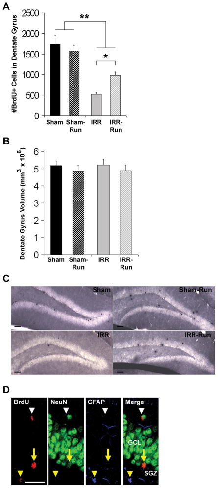
Figure 4.
Daily running partially rescued adult hippocampal neurogenesis months after WBI (Sham, n = 10; Sham-Run, n = 10; IRR, n = 10; IRR-Run, n = 9). All data represent group means. Error bars indicate SEM. A, number of BrdU+ cells in the dentate gyrus at 3 weeks after 5 daily injections of BrdU prior to BM3 training. * p < 0.05; ** main effect of WBI at p < 0.05. B, dentate gyrus volume estimated using the optical fractionator and according to Cavalleri’s principle. C, newly divided cells (peroxidase-stained with anti-BrdU) in the dentate gyrus of Sham, Sham-Run, IRR, and IRR-Run mice. Scale bars indicate 50 μm. Photomicrographs taken at a 10x. D, confocal images of BrdU+ cells in the dentate gyrus that co-expressed NeuN (yellow arrow), GFAP (yellow arrowhead), or neither (white arrow head). Scale bars indicate 25 μm. Confocal images taken at 40x. GCL, granule cell layer. SGZ, subgranular zone.

