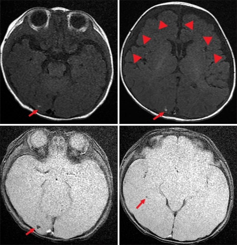
Fig. 2.
Brain MRI of Patient 2 shows multiple small nodular T1-high and gradient echo-dark signal intensity lesions in the right occipital lobe (arrow). This finding is compatible with a calcified inflammatory granuloma as a sequela of previous CMV infection. Also note the subdural effusion and right frontotemporal convexity (arrowheads).

