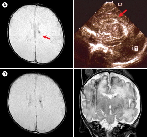
Fig. 3.
(A) Brain images of Patient 4 during the neonatal period. Red arrows point to the small nodular T2-weighted GRE-dark signal intensity lesion in the caudo-thalamic notch on the MRI scan (left) and left germinal matrix hemorrhage on brain sonogram (right). (B) Brain MRI scan of the patient obtained at the diagnosis of thrombocytopenia showed a small nodular T2-weighted axial GRE-(left) and coronal FSE (right)-dark signal intensity lesion in the caudo-thalamic notch (red arrows).

