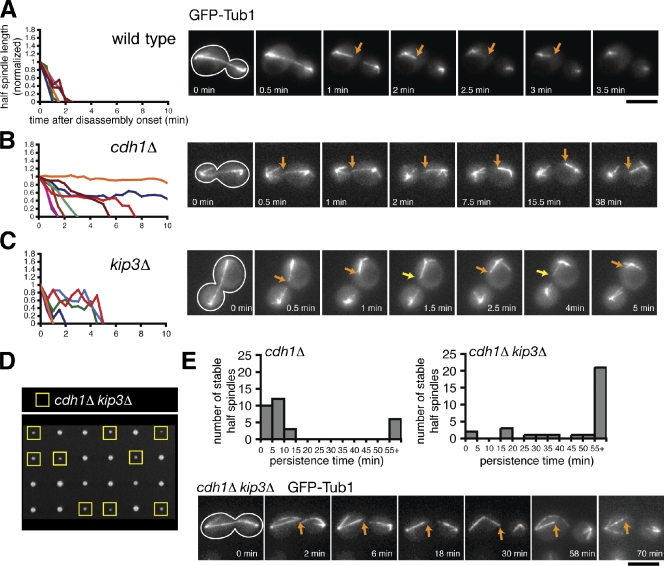
Figure 1.
The combined activities of APCCdh1 and Kip3 are required to depolymerize ipMTs completely. (A–C) Time-lapse fluorescence images of wild-type, cdh1Δ, and kip3Δ cells expressing GFP-Tub1 during mitotic exit. Cell shape is outlined in white. Orange arrows track shrinking spindle halves, whereas the yellow arrows indicate a growing spindle-half. (left) Graphs represent the normalized lengths of depolymerizing spindle halves after the spindle had broken. Each line represents an individual spindle-half that was selected at random. For the cdh1Δ mutant, the shrinkage of four nonhyperstable and four hyperstable spindle halves is charted. Bar, 5 µm. (D) Tetrad dissection of spores resulting from a cross between kip3Δ and cdh1Δ mutants. Offspring possessing both mutations are indicated with yellow boxes. (E) Histograms representing the persistence times of hyperstable spindle halves observed after spindle breakage in cdh1Δ and cdh1Δ kip3Δ mutants (n = 30 for both). (bottom) A series of fluorescence images depicting a cdh1Δ kip3Δ cell after spindle breakage in which the hyperstable half-spindle persisted for at least 70 min.

