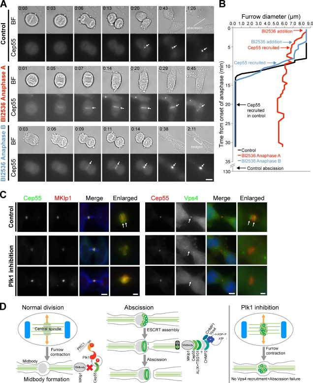
Figure 5.
Plk1 is required for furrow formation and abscission. (A) HeLa cells expressing mCherry-Cep55 were imaged as they exited mitosis after treatment with either DMSO or 1 µM BI2536. Fluorescence (Cep55) and brightfield (BF) still images from the live cell imaging shown in the bottom panels. BI2536 was added as chromosome segregation started in anaphase A or after chromosome segregation, but before furrowing in anaphase B. A dotted line marks abscission in the controls samples. Arrows indicate Cep55 recruitment to the central spindle or midbody. Times are shown in hours:minutes from the start of anaphase, defined as the time point before chromosome segregation was first detected. (B) The graph shows furrow diameter from the onset of anaphase measure in minutes. Arrows show the time of Cep55 recruitment and abscission, if this occurred. (C) HeLa cells treated with DMSO or 1 µM BI2536 for 25 min were stained for DNA with DAPI, mouse anti–α-tubulin (blue), rabbit anti-Cep55 (green), and sheep anti-MKlp1 (red). (right) HeLa cells expressing Vps4-EGFP and Cep55-mCherry were treated with DMSO or BI2536 for 25 min before fixation. Arrows mark the rings of Cep55 and Vps4 flanking the midbody position. (D) A model summarizing how the timely recruitment of Cep55 may be important for proper ESCRT function at the midbody. Bars: (main panels) 5 µm; (enlargements) 1 µm.

