
Figure 2.
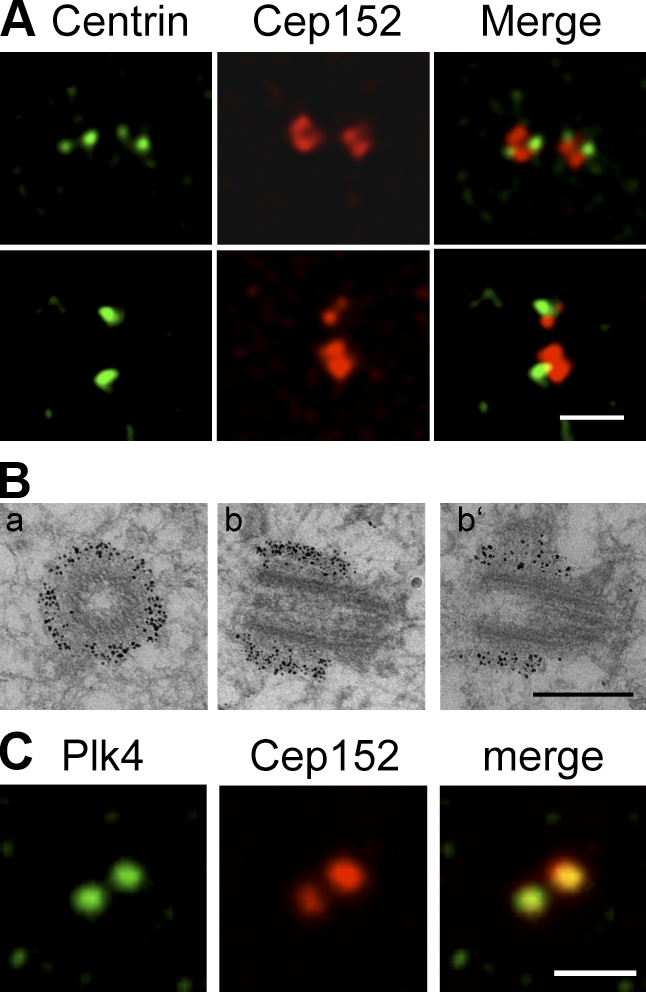
Cep152 localizes to the PCM. (A) Immunofluorescence images showing that a minor fraction of endogenous Cep152 (red) colocalizes with the centriolar marker centrin-2 (green) in U2OS cells. (top) Cell with four centrioles. (bottom) Cell with two centrioles. (B) Immunogold EM of U2OS cells shows that Cep152 localizes to the PCM cloud surrounding the outer wall at the proximal ends of the centriole. Cep152 localization on transversial sections (a) and longitudinal sections of one centriole (b and b’) are shown. (C) Costaining of U2OS cells with antibodies against Cep152 (red) and Plk4 (green). Bars: (A and C) 2 µm; (B) 0.5 µm.