
Figure 2.
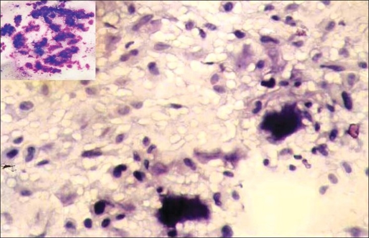
Smear showing polymorphous population of cells dispersed singly along with the matrix. Individual cells are round, oval to spindle with few cells showing binucleation and nuclear indentation (Pap, ×200). Inset: Abundant purple coloured, chondromyxoid matrix (MGG, ×100)