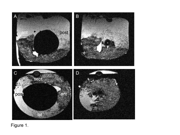
Figure 1.
One hour after reperfusion following a myocardial infarction 4.1 × 10⁷ mesenchymal stem cells labeled with iron nanoparticles were delivered into the mid left anterior descending coronary artery of pigs. (a-d) Sagittal (a, b) and transverse (c, d) views from a post-mortem high-resolution magnetic resonance imaging scan show increased concentrations of iron particles within the mid wall (c) and distal septal (sept) and anterior (ant) walls (d). Lat, lateral wall; post, posterior.

