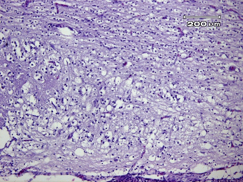
Figure 1.
Embryo of fertile Leghorn egg exposed to Pterocarya fraxinifolia extract. White matter of the spinal cord shows primary malacia and vacuolization (leukomyelomalacia). Large, irregular and empty cavities are affected axons and presumably the result of pooling of myelin and liquefaction necrosis. Spongy appearance is presented (lower part of the figure), in contrast, relatively normal white matter in which myelin sheaths are uniform in diameter (upper part of the figure) (H&E,×100, Bar = 200 µm).

