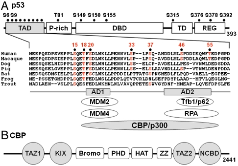
Fig. 1.
Domain organization of p53 and CBP/p300. (A) Schematic of p53 showing TAD (N-terminal transactivation domain), P-rich (proline-rich), DBD (DNA-binding domain), TD (tetramerization domain), and REG (C-terminal regulatory) domains. The sequence alignment of p53 TAD is shown for a few species, and known sites of phosphorylation are indicated by black dots. The AD1 and AD2 motifs are indicated, and proteins that interact directly with them are shown below the sequence alignment. (B) Domains of CBP/p300. The TAZ1 (residues 340–439), KIX (586–672), TAZ2 (1,764–1,855), and NCBD (2,059–2,117) domains interact with p53 TAD and are highlighted in gray.

