Fig. 4.
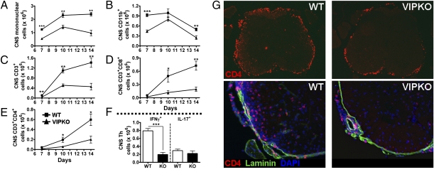
Immune cell infiltration is impaired in VIP KO mice. EAE was induced to WT and VIP KO mice as in Fig. 1 legend, and CNS tissues were collected on days 7, 10, and 14. (A–E) Total numbers of mononuclear cells, as well as CD11b, CD3, CD4, and CD8 cells. (F) Total numbers of Th1 and Th17 cells in the CNS on day 14. The number of infiltrating immune cells was remarkably lower in VIP KO mice (*P < 0.05, **P < 0.01, and ***P < 0.001; Student t test). (G) Photomicrographs at magnifications of 4× (Upper) and 20× (Lower) of spinal cord sections from WT (Left) and VIP KO mice (Right) collected 14 d after immunization and stained by immunofluorescence for CD4 (Alexa 594), laminin (FITC), and DAPI. Thoracic level is shown. Top: CD4 staining only. Lower: Triple staining of laminin, CD4, and DAPI. Note in particular the high abundance of CD4+ cells in the parenchyma of WT but not VIP KO mice (lower two panels), and their apparent nonassociation with laminin-positive blood vessels. Similar results were found at all levels of spinal cords of four sets of WT and VIP KO mice.

