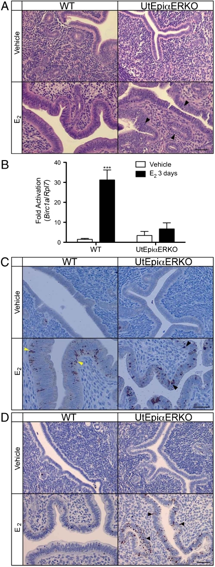
Fig. 5.
Apoptosis-related mediator expressions in WT and UtEpiαERKO uteri. After the treatment of vehicle or E2 for three consecutive days, H&E staining (A) indicated apoptotic appearance of epithelial cells in E2-treated UtEpiαERKO uteri (black arrowheads). Birc1a expression by using real-time PCR analysis (B) increased only in E2-treated WT. Cleaved Caspase-3 IHC (C) increased in both WT (yellow arrowheads) and UtEpiαERKO (black arrowheads) uteri, and TUNEL IHC (D) increased only in E2-treated UtEpiαERKO uteri (black arrowheads) (n = 7). (Scale bar: 20 μm.) Results are mean ± SEM. ***p < 0.001, significantly different from vehicle control of the corresponding genotype, respectively.

