Fig. 1.
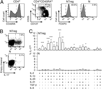
In vitro differentiation of NTreg into IL-17–producing cells. (A) Enriched CD4+ T cells were stained with anti-CD25, -CD45RA, and -CD127 mAb and CD45RA+ cells (left histogram) were sorted by flow cytometry into naive conventional CD4+ T cells (N, CD25−CD127high) and naive Treg (NTreg, CD25+CD127low) (dot plot). A fraction of sorted populations was stained with anti-FOXP3 mAb and analyzed by flow cytometry (right histograms). (B and C) Sorted N and NTreg were stimulated in vitro with anti-CD2/CD3/CD28 microbeads in the presence of the indicated cytokines and cultured for 12 d in the absence or presence of IL-2, as indicated. IL-17 production was assessed by intracellular cytokine staining and flow cytometry analysis following stimulation with PMA/ionomycin. Dot plots for N and NTreg from one donor stimulated in the presence of IL-2, IL-1β, IL-23, and TGF-β are shown in B. Results for all conditions and donors are shown in C (mean ± SD, n = 6). Statistical analyses were performed using two-tailed t test. *P < 0.05; **P < 0.01; ***P < 0.001.

