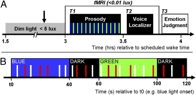
Fig. 1.
Experimental design. (A) General protocol. Arrow indicates pupil dilator administration. Time relative to scheduled wake time (h). T1 (task 1): first fMRI task, consisting of a gender discrimination of auditory vocalizations while exposed to alternating blue and green monochromatic ambient light (see B for details). T2 (task 2): second fMRI task (voice localizer); its main aim was to identify the voice-sensitive area of the temporal cortex. Participants performed a 1-back task with the voice stimuli from task 1 (anger and neutral pseudoword) and nonvoice white-noise auditory stimuli replicating the envelope (EN) or the mean of the fundamental (F0) of the original voice stimuli from task 1. T3 (task 3): emotional judgment task performed outside the MRI scanner, in which the emotions of all of the auditory stimuli presented in T1 were evaluated by the participants on a five-item Likert scale. (B) Detailed experimental procedures of the gender discrimination task (T1). Time (s) relative to t0, a time point arbitrarily chosen as a blue light onset of the session. Monochromatic [blue (473 nm) or green (427 nm)] ambient light exposures lasted 40 s and were separated by 15- to 25-s periods of darkness (mean duration, 20 s). Anger (red bars) and neutral (white bars) prosody vocalizations (meaningless word-like sounds; half neutral, half anger) were pseudorandomly and evenly administered in each light condition throughout the entire session (interstimuli interval, 3–11 s; mean, 4.8 s).

