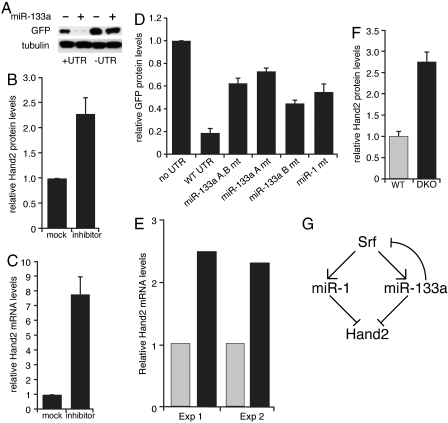
Fig. 5.
miR-133a regulation of Hand2 expression. (A) Western blot demonstrating that addition of miR-133a oligonucleotides to nondifferentiated C2C12 cells only inhibits GFP expression from reporter constructs containing the Hand2 3′UTR. Similar results were observed in three independent experiments. (B) miR-133a-blocking oligonucleotides increase endogenous Hand2 protein levels in primary cardiomyocytes. Hand2 protein levels were normalized to tubulin. (Mean ± SE are shown.) (C) miR-133a-blocking oligonucleotides increase Hand2 mRNA levels in primary cardiomyocytes, as determined by quantitative RT-PCR. Results shown represent three independent experiments. (Mean ± SE are shown.) (D) Mutation of miR-133a MREs relieves reporter repression in differentiated C2C12 cells. Effect of mutating the miR-1 MRE is shown for comparison. GFP protein levels quantified by densitometry were normalized to tubulin. Results represent six independent experiments. (Mean ± SE are shown.) (E) RT-PCR analysis of Hand2 mRNA levels in heart samples from miR-133a double knockout (DKO) mice and wild-type littermates. Five hearts at postnatal day 1 from each condition were used for each experiment. Gray bars represent wild type, black bars represent double knockouts. (F) Hand2 protein levels, relative to tubulin, in hearts from miR-133a DKO and wild-type littermates. Hearts from three wild-type and miR-133a DKO mice were analyzed. (Mean ± SE are shown.) (G) Model for miR-1 and miR-133a regulation of Srf and Hand2 expression, wherein partial loss of miR-133a feedback inhibition of Srf augments miR-1 response.

