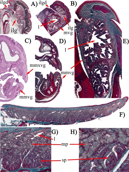
Fig. 15.
A, Masson's trichrome-stained transverse histology section of S. crocodilurus showing the mixed seromucous lobules. B, Masson's trichrome-stained transverse histology section of V. varius showing the intratubular lumina feeding into the large central lumen of the mandibular venom gland and the individual mucous lobules dorsally positioned that collectively make up the gland. C, periodic acid-Schiff-stained transverse histology section of O. apodus revealing that small mandibular mixed type venom gland and small maxillary mixed type venom gland arrangements are present, each with a central lumen. Masson's trichrome-stained histology sections of the G. infernalis mixed mandibular venom gland show the intratubular lumina that feed into the large central lumen (D) and longitudinal (E) and transverse sections (F, G, and H). ilg, infralabial gland; ilgd, infralabial gland duct; l, lumen; mmvg, mixed mandibular venom gland; mmxvg, mixed maxillary venom gland; mp, mucous part; mvg, mandibular venom gland; sp, serous part.

