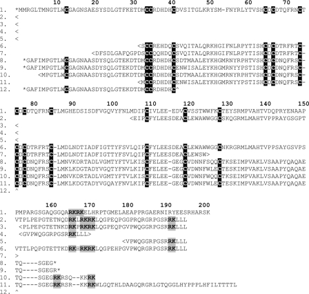
Fig. 6.
Sequence alignment of phospholipase A2 (type III) toxin precursors from mandibular venom glands. 1, C. warreni (GU441524); 2, V. gilleni (GU441524); 3, V. scalaris (GU441526); 4, V. glauerti (GU441527); 5, V. tristis (GU441528); 6, V. komodoensis (B6CJU9); 7, V. varius (Q2XXL5); 8, H. suspectum (P80003); 9, H. suspectum (P16354); 10, H. suspectum (EU790967); 11, H. suspectum (EU790968); 12, H. horridum (P04362). Cleavage motifs are highlighted in gray, cysteines are in black, and the signal sequence is shown in lowercase. < and > designate incomplete N and C termini of the precursor, respectively. * designates the N and C termini of a post-translationally processed protein sequence. ∧ designates a protein sequencing fragment with an incomplete C terminus.

