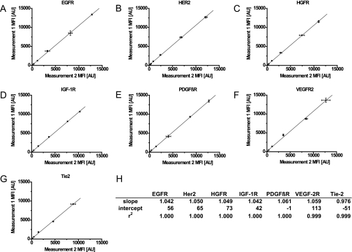
Fig. 3.
Repetitive measurements of seven receptor tyrosine kinases from same sample. The diagrams show the results of two sequentially conducted multiplex sandwich immunoassays for the EGFR (A), HER2 (B), HGFR (C), IGF-1R (D), PDGFRβ (E), VEGFR2 (F), and Tie-2 (G) using the same dilution series. Antibody-coupled beads were incubated with recombinant standards at seven different 4-fold dilutions starting for EGFR, HER2, and PDGFβR at 20,000 pg/ml; for IGF-1R and VEGFR at 100,000 pg/ml; for HGFR at 50,000 pg/ml; and for Tie-2 at 10,000 pg/ml. A mixture of biotinylated antibodies specific for the respective RTK, together with SAPE, was used to detect the analytes in the readout system (Luminex 100). The median fluorescence intensities (MFI) of the two sequential assays were plotted against each other and fitted linearly. Standard deviations were calculated from three technical replicates. The table (H) contains slopes, intercepts, and correlation coefficients of the fits for the seven assays. The calculated correlations and slopes close to 1.000 confirm the hypothesis that the analyte can be analyzed twice without changing the concentration of the analyte itself. AU, arbitrary units.

