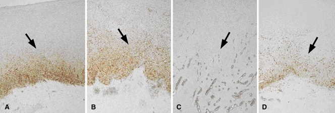
Figure 3:
HSP production following combined RF ablation and liposomal chemotherapy. A–D, Photomicrographs show rim staining of HSP70 (arrows) surrounding zone of coagulation on sections from tumors excised at 24 hours. (Original magnification, ×4.) A, RF ablation alone. B, RF ablation–doxorubicin. C, Paclitaxel–RF ablation. D, Paclitaxel–RF ablation–doxorubicin. HSP70 staining was substantially diminished in tumors treated with paclitaxel–RF ablation, with a comparatively less intensely positive rim observed for paclitaxel–RF ablation–doxorubicin.

