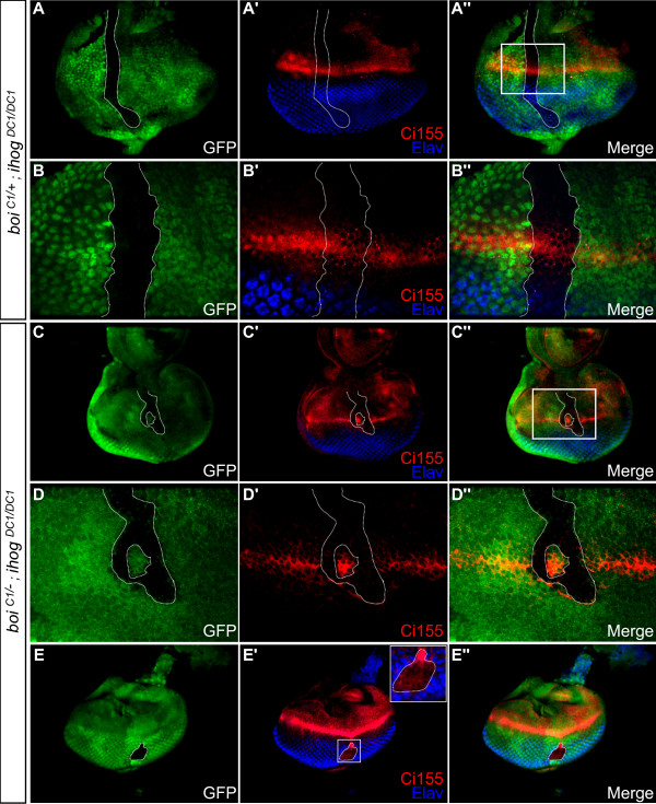
Figure 3.
Cells mutant for both ihog and boi do not activate the Hh pathway, disrupting R-cell differentiation in the developing eye disc. (A-B'') Control in which an ihogDC1/DC1 clone (green fluorescent protein (GFP)-negative cells marked by dotted line) was generated in an eye disc of a boiC1/+ heterozygote. (A-A'') Low magnification view of entire disc. (B-B'') High magnification view of boxed area in (A''). Control clones show normal accumulation of cytoplasmic Ci155 (red) near the MF, and normal expression of Elav (blue) among differentiating R-cells posterior to the MF. (C-E'') An ihogDC1/DC1 clone (dotted line) in a boiC1/- mutant. (C-C'') Low magnification view. (D-D'') Higher magnification view of boxed area in (C''). Ci155 expression (red) is undetectable in cells mutant for both ihog and boi (GFP-negative, dotted line). (E-E'') In a double mutant clone situated well posterior to the MF, the absence of Elav and the ectopic expression of Ci155 indicate a delay of R-cell differentiation. Anterior is at the top in all panels.

