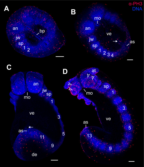
Figure 4.
Cell division patterns in segmenting embryos of the ovoviviparous onychophoran Euperipatoides rowelli (Peripatopsidae). The full number of 15 leg-bearing segments has not been established yet. Double-labelling with the DNA-selective dye Hoechst (Bisbenzimide, blue) and anti-phospho-histone H3 antibody (α-PH3, red). (A) Flat preparation of an embryo with two leg-bearing segments. (B) Flat preparation of an embryo with four leg-bearing segments. (C) Ventrolateral view of a late stage 3 embryo with 11 leg-bearing segments. (D) Ventrolateral view of an early stage 4 embryo with 13 leg-bearing segments. Note the concentric rings of proliferating cells in the antennal segment (arrowheads). Arrows (in B and C) point to remnants of the blastopore in front of the future anus. Leg-bearing segments numbered. Abbreviations: an, antennal segment; as, anus; at, presumptive antenna; bp, blastopore; de, dorsal extra-embryonic ectoderm; jw, jaw segment/presumptive jaw; mo, embryonic mouth; sp, slime papilla segment/presumptive slime papilla; ve, ventral extra-embryonic ectoderm. Scale bars: A-D, 200 μm.

