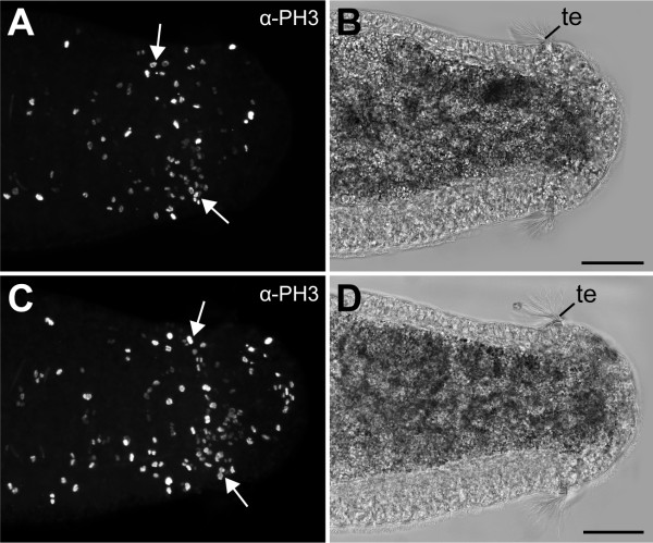
Figure 7.
Cell division pattern at the posterior end in larvae of the polychaetous annelid Capitella teleta (Scolecida, Capitellidae). Confocal (A, C) and light micrographs (B, D) of posterior ends of an early (A, B) and a late stage 8 larvae (C, D), labelled with an α-PH3 antibody. Note a localised region of high cell proliferation (arrows) in front of the telotroch (te). Scale bars: 50 μm.

